

Translate this page into:
Ectodermal dysplasia: A case report
-
Received: ,
Accepted: ,
How to cite this article: Rishi S, Arora B, Dubey D, Chawla P. Ectodermal dysplasia: A case report. Adesh Univ J Med Sci Res 2022;4:105-8.
Abstract
Ectodermal dysplasia (ED) is a rare hereditary disorder which is characterized by the defect of tissue of ectodermal origin. It is heterogeneous group of hereditary disorder which affects approximately one in every 100,000 births. Children who have ED may have various manifestations which may or may not involve teeth, skin, hair, nails, and sweat glands. Here, we present one such case of ectodermal dysplasia in 12-year-old patient.
Keywords
Anodontia
Hypohidrosis
Xerostomia

INTRODUCTION
Ectodermal dysplasia (ED) is rare hereditary disorder which is congenital, diffuse and non- progressive. It is a primary developmental defect of two or more tissues derived from the ectoderm. Tissues which are primarily affected are skin, hair, nails, eccrine glands, and teeth.[1,2] The ED is manifested by defects only in the ectoderm structure along with other anomalies. The most common ED is hypohidrotic and hidrotic ED which is inherited as an X-linked recessive trait.
Several defects which include hypohidrosis, anomalous dentition, onychodysplasia, and hypotrichosis are seen in ED.
Facial appearance is typical which is characterized by frontal bossing, sunken cheeks, a saddle nose, and hyperpigmented skin around the eyes and low-set ears. Dental manifestations include conical or peg shape teeth, hypodontia or anodontia, and delayed eruption of permanent teeth. In hypohydrotic ED eccrine sweat glands may be absent or sparse and rudimentary.[3]
CASE REPORT
A 12-year-old male patient came to department of Pedodontics and Preventive Dentistry at Adesh Institute of Dental Sciences and Research Bathinda with his father, with the chief complaint of multiple missing teeth in relation to the upper and lower arches. There was no history of birth complication during his delivery. His father gave history of fine hair growth, dryness of mouth, decreased sweating and reduced lacrimation.
His father also gave a history of frequent bouts of fever throughout childhood, and also mentioned that his boy was intolerant to heat.
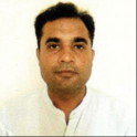
On extraoral examination, he had frontal bossing, a prominent supraorbital ridge, sunken cheeks, thick lower lip, sparse hair, scanty eyebrows, and brittle nails [Figure 1a-d].

- (a-c) Extraoral photograph showing frontal bossing, prominent supraorbital ridge, thick lower lip, sparse hair, sunken cheeks, and scanty eyebrows, (d) photograph showing brittle nails.
On intraoral examination, only seven permanent teeth were present which include maxillary right central incisor, first molar, left maxillary central incisor and first molar, right mandibular first molar and mandibular left canine and first molar [Figure 2a-c]. OPG was taken and it revealed multiple missing tooth buds with unerupted tooth including maxillary right canine and second premolar, maxillary left canine, first and second premolar, mandibular right canine and first premolar and left first premolar [Figure 3]. On the basis of history, clinical and radiographic examination, ED was diagnosed. Patient was advised to consume water frequently to maintain proper hydration and artificial salivary substitutes were advised to reduce Xerostomia. Patient was advised for dental implants in future, after the completion of dentofacial growth. Patient was also referred to ophthalmologist for reduced lacrimation.

- (a-c) Intraoral photograph showing seven permanent teeth were present- maxillary right central incisor, first molar, left maxillary central incisor and first molar, right mandibular first molar, and mandibular left canine and first molar.

- Panoramic radiograph showing multiple missing tooth buds with unerupted tooth buds including maxillary right canine and second premolar, maxillary left canine, first and second premolar, mandibular right canine and first premolar, and left first premolar.
DISCUSSION
ED is a group of hereditary disorders characterized by developmental abnormalities of ectodermal derivative structure.
Etiology and pathogenesis
ED occurs due to the defects of the genes regulating epithelial and mesenchymal signaling. The defective genes identified are ED-1, muscle segment homeobox homology-1, ectodysplasia-1 (EDAR), paired box genes and human homolog of mouse dl, EDARDD, and WNT 10A. The genes such as ED, EDAR, and others collectively act to produce protein ectodysplasin-1. This protein is essential in the interaction between ectoderm and mesoderm, which occurs at 6th week of intrauterine life. Ectoderm-mesoderm interactions are essential for the formation of several structures that arise from the ectoderm, including the skin, hair, nails, teeth, and sweat glands. Hence, any mutation at this point leads to severe form of ED. ED1 mutations are observed in X-linked recessive form, and EDAR, EDARDD, and WNT 10A mutations are observed in autosomal dominant and recessive inheritance.
First classification of ED was proposed by Freire-Maia and Pinheiro in 1982.
According to them ED is classified into different subgroups
ED1: Presence or absence of hair anomalies or trichodysplasia
ED2: Dental anomalies
ED3: Nail abnormalities or onychodysplasia
ED4: Eccrine dysfunction or dyshidrosis.
ED is further categorized into:
Group A disorder which involves defect of at least two ectodermal structure as mentioned above, with or without defects of other structure.
Group B disorder which involves one of the classical ectodermal structure including hair, nail, teeth, or eccrine glands in combination with the defect in any of the ectodermal structure (ear, lip).
Extraoral features include fine, sparse hair over the scalp along with extensive scaling of the skin and unexplained pyrexia along with heat intolerance is seen, most commonly occurs due to anhidrosis. Normal intelligence is observed. Other extraoral features are frontal bossing, sunken cheeks, depressed nasal bridge, thick everted protuberant lips, wrinkled or hyperpigmented periorbital skin, and a large low set of ears.[4] Intraoral features include, missing permanent teeth and teeth which are most commonly present are the maxillary central incisors and canines present with a conical crown form. In rare instances, one or both jaws may be edentulous and the alveolar processes may not develop due to the absence of teeth.[5] Patient who reported here had the involvement of hair, sweat glands, multiple missing permanent teeth and nail dystrophy. Diagnosis of ectodermal dysplasia is based on family history, clinical history, and radiographic examination.[6]
Diagnosis
Diagnosis is based on the episodes of hyperpyrexia, lack or sparse type of hair, absence of teeth, and tooth buds and tooth morphology. Peeling of the skin at birth, eczema, asthma, and frequent respiratory infections may be additional clues. However, during early infancy diagnosis is difficult because manifestations involving teeth, hair, and inability to sweat are hard to detect. However, attempts have been made in past to develop objective diagnostic criteria based on number and distribution of sweat pores and amount of sweat produced. The structural and biochemical characteristics of hair have also been studied.[7]
Other diagnostic criteria have been dermatoglyphic analysis, characteristics of lacrimal secretion, and distribution and pattern of scalp hair. Missing teeth can also be an important sign.[8]
Treatment of ED depend on which ectodermal structure is involved. In our case, temperature of body was slightly elevated which is associated with xerostomia. Hence, patient is advised to have frequent consumption of water to maintain hydration and thermoregulation and artificial salivary substitute is advised for xerostomia. Early dental evaluation and intervention is advised for patients with dental defects. The key of success for management of ED is quick diagnosis and prosthetic rehabilitation by the multidisciplinary approach.[9] Pedodontist play an important role for the successful management of ED because they are better trained in child psychological and behavioral management.[10]
It is also important to instill awareness among parents regarding early management. To ensure adequate care, child with ED should be managed by a team which includes pediatrician, pediatric dentist, prosthodontist, dermatologist, otolaryngologist, speech therapist, and psychologist. However, an appropriate change has to be made in the denture so that the appearance is always appropriate for the age. Relining or change of dentures should be done every 1 or 2 years due to preadolescent growth in jaw dimensions, wear of the acrylic teeth, under extension of the dentures and posterior open bite. Cephalometric analysis has shown favorable growth of maxilla and mandible following the placement of dentures.[11]
Implants are not recommended in children before skeletal maturity. However, the successful use of any prosthesis depends on the cooperation and communication between members of dental team and the patient.
CONCLUSION
ED is a rare genetic disorder which has many overlapping features and leading to difficulty in classification. The clinical manifestations of ED cause significant social problems in affected individuals. Prosthodontist and pedodontist play an important role for the management of this.[8] Prognosis of ED is good and the patients of ectodermal dysplasia have normal life span.
Declaration of patient consent
The authors certify that they have obtained all appropriate patient consent.
Conflicts of interest
There are no conflict of interest.
Financial support and sponsorship
Nil.
References
- Textbook of Oral Diagnosis In: Examination of Teeth (4th ed). Saint Louis: Mosby; 1974. p. :201.
- [Google Scholar]
- Hydrotic or hypohydrotic ectodermal dysplasia: Diagnostic dilemmas (case report) Int J Curr Microbiol Appl Sci. 2015;4:778-83.
- [Google Scholar]
- Ectodermal dysplasia (ED) syndrome: Case report. Biomedicine (Taipei). 2014;4:27.
- [CrossRef] [PubMed] [Google Scholar]
- Rapp-Hodgkin syndrome: An ectodermal dysplasia involving the teeth, hair, nails and palate. Oral Surg Oral Med Oral Pathol. 1989;67:50-62.
- [CrossRef] [PubMed] [Google Scholar]
- Hypohydrotic ectodermal dysplasia: A case report and review. Int J Adv Health Sci. 2014;1:38-41.
- [Google Scholar]
- Hypohidrotic ectodermal dysplasia with hypothyroidism. J Pediatr. 1981;98:223-7.
- [CrossRef] [PubMed] [Google Scholar]
- Hypohidrotic ectodermal dysplasia: Dental, clinical, genetic and dermatoglyphic findingsof three cases. J Clin Pediatr Dent. 2001;26:5-12.
- [CrossRef] [PubMed] [Google Scholar]
- Ectodermal dysplasia with oligodontia: A rare case-rehabilitation by prosthetic management. Acta Sci Dent Sci. 2019;3:12-4.
- [CrossRef] [Google Scholar]
- Hereditary ectodermal dysplasia: A case report. J Indian Soc Pedod Prev Dent. 2002;20:37-40.
- [Google Scholar]


